The Advanced Electron Microscopy (AEM) facility is a shared resource of the BSD, managed by the Office of Shared Research Facilities (OSRF). First developed by Ernst Ruska in the 1930’s, the principles of electron microscopy (EM) are very similar to light microscopy (LM), but instead of using photons and glass lenses, electrons are accelerated through a vacuum and electromagnetic lenses focus them onto biological samples, allowing researchers to collect images of proteins, cells, and tissues.
Even though EM historically has been foundational in both cell biology and structural biology, it took many decades of developments for Ruska’s original vison to be completely realized. In 2017, the Nobel prize in Chemistry was awarded for the development of cryo-EM, which involves flash-freezing solutions before imaging (this technology was recently used in the world-wide effort to determine the COVID virus’s spike protein). This kick-started the “Resolution Revolution” with increasingly powerful microscopes, which UChicago quickly joined as well.
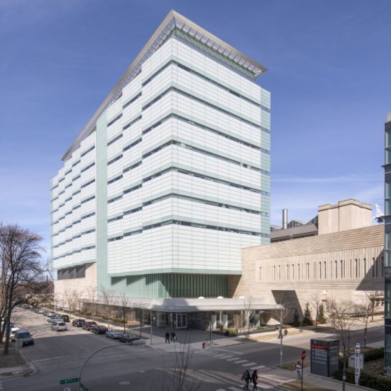
In 2017, the University already had an EM facility dedicated to cryo-preservation and 3D tomography in the sub-basement of the Gordon Center for Integrative Science, but the facility needed additional space to accommodate a new 12-foot tall cryo-EM called the Titan Krios. The EM facility team went on a search for space across campus, discovering a new research home while unearthing an interesting bit of history in the Franklin McLean Institute (FMI).
The FMI building was originally built in 1953 and was known as Argonne Cancer Research Hospital until 1973. It was one of the first institutions equipped to use radiation sources for studying of cancer, and housed the university’s original cyclotron, a type of particle accelerator that produces radioactive isotopes that can be used for imaging procedures, installed in 1968.
After the cyclotron and other equipment in the Cancer Research Hospital was decommissioned in the 1990s, the space was converted to offices, and it would have stayed that way if not for the need to find a home for the Titan Krios. In 2018, construction of the new AEM facility started by fully gutting the space and partially removing a 3-foot-thick concrete, lead, and iron wall that had been shielding for a linear accelerator. The newly created space not only had room for the Titan Krios, but also three other EMs, an operator’s room, two offices, and a mezzanine level with supporting equipment necessary for all the scopes.