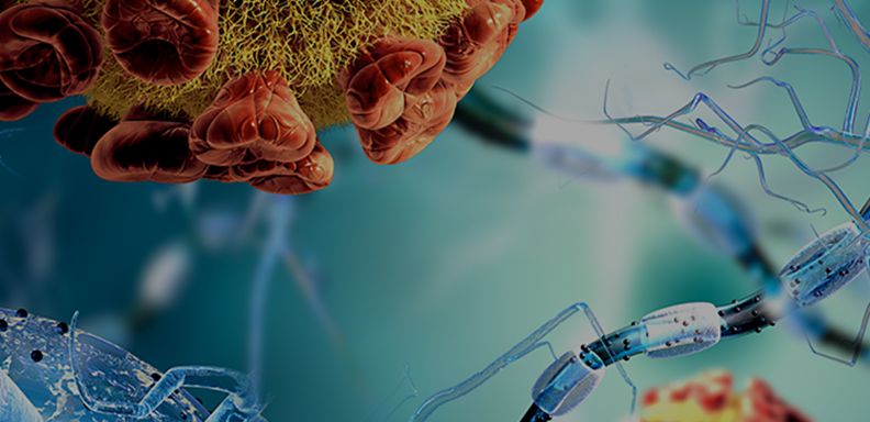
Actin filaments form the cytoskeleton – or structural features – of cells in the body. These filaments undergo various types of strain throughout their lives, being pulled, pushed and otherwise stressed by forces both inside and outside of the cell. This tension can lead to damage to the filaments. When the damage is not repaired, it may result in health issues as varied as hearing loss and cardiac abnormalities, depending on the type of cell affected.
A group of researchers at the University of Chicago set out to use computer simulation to better understand what happens to actin filaments under tension and how they recruit proteins to repair damage when it occurs. Their findings, recently published online by Biophysical Journal in the paper “Cracked actin filaments as mechanosensitive receptors,” reveal a previously unknown binding site that is exposed when filaments are damaged. Proteins travel to that site to repair the damage.
The team and co-authors include Margaret Gardel, PhD, Horace B. Horton Professor of Physics, Molecular Genetics and Cell Biology and Molecular Engineering; David Kovar, PhD, Professor of Molecular Genetics & Cell Biology and Biochemistry & Molecular Biology; Gregory Voth, PhD, Haig P. Papazian Distinguished Service Professor of Chemistry, the James Franck Institute, and the Institute for Biophysical Dynamics; and Vilmos Zsolnay, PhD ’23, a former student in UChicago’s Graduate Program in the Biophysical Sciences.
“This discovery has made us completely rethink what we thought we knew about the fundamental properties of actin filaments,” Kovar said. “We were previously aware of three binding sites, but we have now uncovered a fourth that becomes exposed when tension causes a crack in the actin filament. This new binding site could change what proteins bind to the structures and how they bind to them.”
“This work not only illuminates the structural changes that occur in actin filaments under mechanical tension, but it also has implications for our understanding of the development and treatment of various pathologies,” Gardel said.
Predicting the behavior of proteins
The team used all-atom molecular dynamics simulations to investigate how tension alters the actin filament binding surface, and protein-protein docking simulations to explore interactions with associated proteins. These complex computational models use the known physics of atoms to predict how proteins and other molecular systems behave under specific circumstances.
The molecular dynamics simulations showed that actin filaments, which look like two strings of pearls wrapped around each other in a helix, do not see a distortion or separation of their individual “pearls” when tension is applied or fragment simultaneously along both “strings,” as was theorized. Instead, a fissure or crack first appears in only one of the strings. This crack exposes a set of “cryptic,” or buried, binding sites that can recruit proteins.
The researchers then used protein-protein docking simulations to see if a particular family of proteins called LIM domains would bind to the crack in the filament. Previous wet lab experiments showed LIM domain proteins binding to actin filaments under strain, but not where they were binding. The model established that LIM domain proteins overwhelming prefer to bind to the cryptic binding sites to stabilize the filament, indicating that they are critical to repairing the structures.
The necessity of interdisciplinarity
The simulations were performed and analyzed by Zsolnay, the paper’s first author and current consultant at Bain & Company. The UChicago biophysical sciences program he graduated from requires that students conduct their research under the mentorship of two professors – typically one in the biological sciences and the other in the physical sciences – and to explore the science at the interface between the disciplines. Zsolnay worked in both Kovar’s lab and Voth’s lab.
The three labs involved in this research have a history of collaboration, including a multi-year Department of Defense MURI (Multidisciplinary University Research Initiative) grant awarded in 2015 to explore how biological systems sense and respond to mechanical force. The grant helped support this project. This interdisciplinarity is a hallmark of research at the University – and one of its biggest strengths, said Voth.
“This research couldn’t have been done without the expertise of our three labs,” Voth said. “The University is known for fostering these types of collaborations and for innovative programs and institutes like the Biophysical Sciences graduate program and the Institute for Biophysical Dynamics where interdisciplinarity is a key feature. The future of scientific research is really at these interfaces between the disciplines.”
The researchers now plan to conduct in vitro experiments to further explore the behavior of actin filaments under strain and to uncover when and why filaments may fail to repair themselves.


