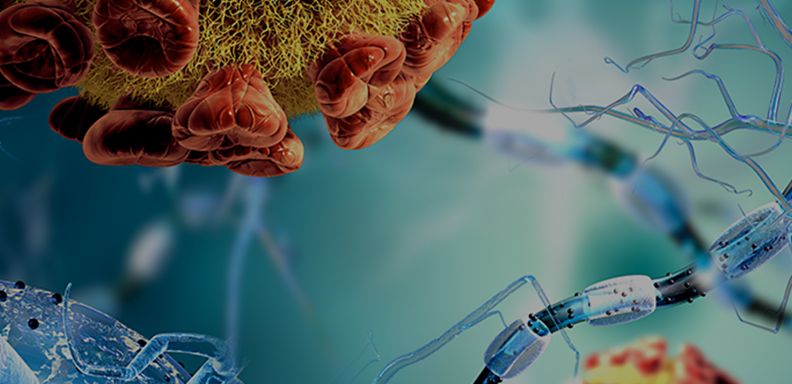
Over the course of evolutionary history, cooperation between single-celled organisms has formed the foundation of complex cellular networks, leading to multicellular life. The physical organization of multicellular organisms is essential for complex life and is largely enabled by a cell’s ability to adhere to others. One example of this is when neurons need to find their targets in order for complex brain circuits to form.
While there are various cell surface proteins that play a role in cell adhesion, Teneurins, a group of cell-surface receptors that are evolutionarily conserved in many organisms, are of great interest. Teneurins are crucial for embryonic development and they play roles in helping neurons form proper connections. Mutations in teneurins are linked to the progression of certain cancers, such as glioblastomas and ovarian cancers, and they are also linked to microphthalmia and congenital anosmia, two developmental birth defects.
Generally, proteins have certain regions that give rise to special functions. Teneurins possess a unique ability to self-associate with other teneurin proteins from the same cell (cis) and also from a different cell (trans). A protein bound to one other protein is called a dimer and a protein bound to the same type of protein is called a homodimer. Currently, it is not known what functions self-associating teneurins play.
In a new study, Jiangxian Li, PhD, and Sumit Bandekar, PhD, postdoctoral scholars in the lab of Demet Araç, PhD, Associate Professor of Biochemistry and Molecular Biology at the University of Chicago, used the fruit fly version of teneurin, called Ten-m, to explain the molecular mechanisms behind teneurin self-association. Using protein crystallography, the researchers revealed that association between teneurins is asymmetric and employs the transthyretin-related domain and the β-propeller domain respectively. When this interface is repeated, it can build teneurin complexes larger than dimers, compatible with both cis and trans configurations. These long teneurin assemblies have the potential to form tight structures reminiscent of zippers and “zip” two cells together.
Along with determining the first invertebrate teneurin crystal structure, Li and Bandekar also created mutants of Ten-m that lose the ability to dimerize, so they can study the functions of teneurin dimers and ultimately the roles teneurins play in development and axon guidance. “What these [mutants] allow us to do is to test the biological function of the dimer without affecting other aspects of the protein,” they said.
The study has shed light on the mechanism behind teneurin dimizeration and provides a gateway to studying the functions of these essential proteins. In addition, Li and Bandekar have established targets for small molecular inhibitors for potential drug applications in teneurin-related diseases.
The study, “The structure of fly Teneurin-m reveals an asymmetric self-assembly that allows expansion into zippers,” was published in EMBO Reports on May 11, 2023. The research was supported by the National Cancer Institute, the National Institute of General Medical Sciences, the National Institutes of Health, and the Department of Energy. Disclaimer: This content is solely the responsibility of the authors and does not necessarily reflect the official view of the National Institutes of Health.


