Scientists at the University of Chicago and the MRC Laboratory of Molecular Biology (Cambridge, England) have teamed up to better understand how membrane proteins are made.
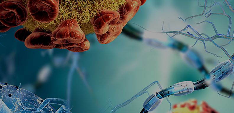
Human cells contain thousands of different types of these proteins, which must fold up within membranes to perform their crucial functions, including as receptors, transporters and energy producing machines. When these proteins are made incorrectly, it can lead to a number of diseases, including cystic fibrosis, retinitis pigmentosa, and Charcot-Marie Tooth disease.
“Researchers have been studying the biosynthesis of membrane proteins for decades,” said Robert Keenan, PhD, a Professor of Biochemistry and Molecular Biophysics at UChicago and co-lead on the new project. “We have a pretty good idea of how the cell makes the simplest types of membrane proteins, but how this works for more elaborate membrane proteins—those with multiple transmembrane segments—has been less clear. It was generally assumed that the process for making multipass proteins would be very similar, but that idea didn’t always fit with observations.”
Several years ago, researchers in the Keenan lab discovered a set of proteins that seemed important for making multipass proteins. Since then, working in collaboration with Ramanujan Hegde’s group at the MRC Laboratory of Molecular Biology, the researchers have focused on understanding what these components do and how they work.
Results of this research have been published online by Nature in a pair of papers, “Substrate-driven assembly of a translocon for multipass membrane proteins” and “Mechanism of an intramembrane chaperone for multipass membrane proteins.”
A complex team, working together
Most membrane proteins are made by ribosomes on a part of the cell known as the endoplasmic reticulum (ER). As the new protein is being made, it enters the ER membrane where it folds into the correct shape to serve its function.
The machinery that coordinates the insertion of the new proteins into the membrane is called a “translocon.” This machine is built around a protein complex called Sec61 that can move portions of the new protein into or across the membrane. When a multipass protein is being made, a number of other components are recruited, forming what the researchers call the “multipass translocon.”
The researchers’ first paper, led by the Keenan group, details a series of experiments in cells and in vitro that reveal how and when the multipass translocon assembles, and what can go wrong when multipass proteins are made in its absence. The striking thing about the translocon, the researchers say, is that it adds and subtracts components as needed while the protein is being made.
“What we show is that the translocon is dynamic. As a new protein begins to engage with the machinery in the membrane, it recruits different factors that come and go at different points in the synthesis, depending on what the protein needs,” Keenan said. “For multipass proteins, this remodeling process is important to help them fold into their final functional forms.”
In the second paper, led by the Hegde group, the scientists took a closer look at a component of the mutlipass translocon known as the PAT complex. Here they showed how the PAT complex acts as a “chaperone” to protect multipass proteins in the membrane, so they can more easily fold into the correct shape. This work also suggests how portions of a multipass protein can be inserted into the membrane through the multipass components, rather than through Sec61 as was previously thought.
These studies alter the conventional view of how multipass proteins are made and provide a framework for understanding how the actions of other biogenesis factors are coordinated at the translocon during the synthesis of different types of proteins.
“We are fortunate to work with such a terrific group of young scientists whose talent and dedication made this research possible,” Keenan said.
Creating a complete snapshot
The teams plan to continue their research to generate a more complete picture of multipass protein biogenesis.
“Ultimately, we'd like to have a molecular movie of a protein folding at the multipass translocon. That will require a series of structural snapshots of folding intermediates, from early stages with just a few transmembrane segments bound to the machinery, to later stages where nearly all of the transmembrane segments are present,” Keenan said. “From a technical standpoint, this is very challenging, but it’s an important goal.”
A better understanding of how cells make multipass proteins could someday lead to new treatments for human diseases, he said.
Additional authors on the papers include Arunkumar Sundaram, Melvin Yamsek and Frank Zhong of the University of Chicago; and Yogesh Hooda, Ramanujan S. Hedge, Luka Smalinskaite, Min Kyung Kim, and Aaron J. Lewis of Cambridge University.
This work was funded by the National Science Foundation, the UK Medical Research Council, and the National Institute of General Medical Sciences of the National Institutes of Health.


